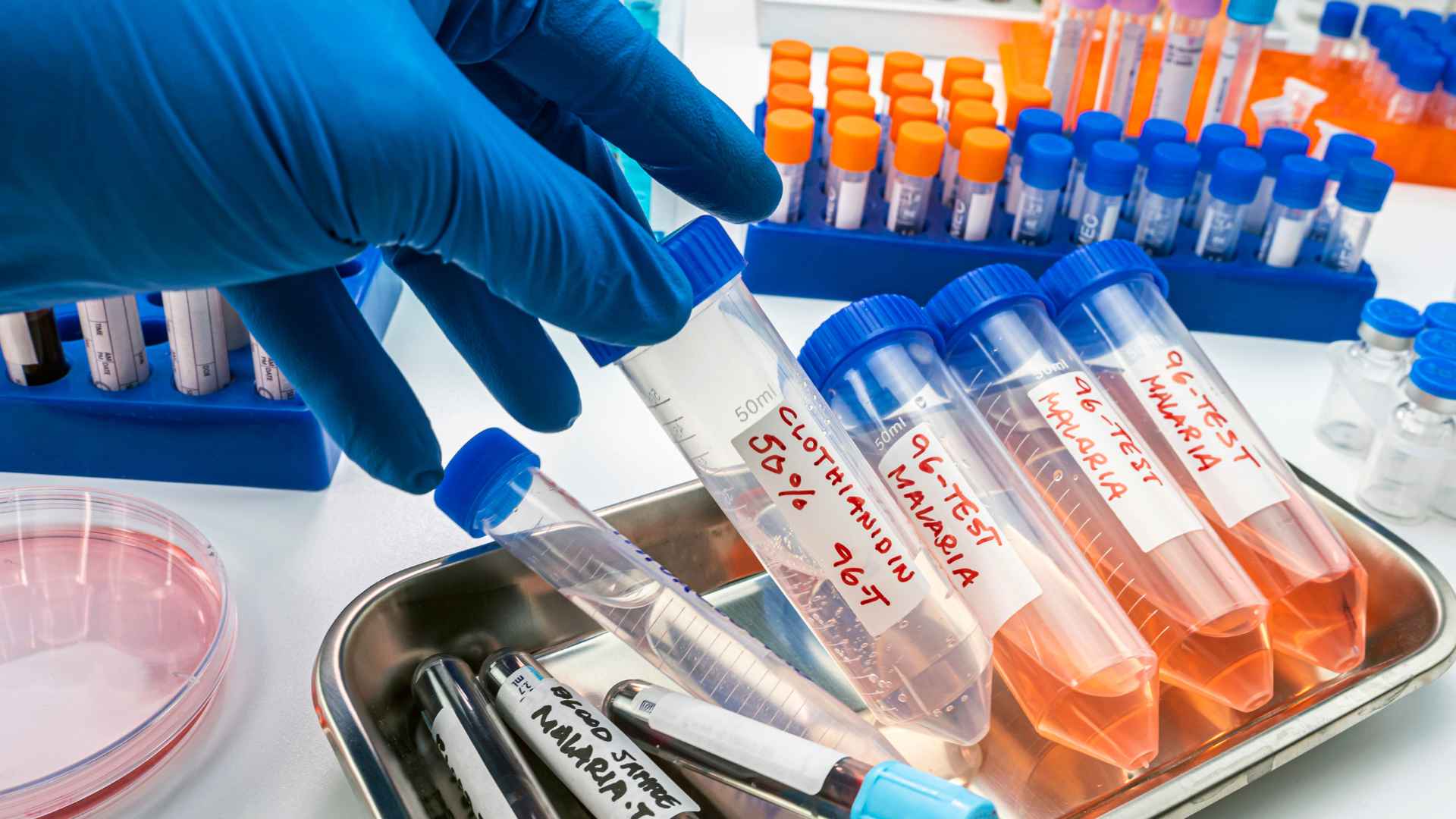
you’re standing at the intersection of art and science, brush in hand—not to paint a canvas, but to reveal secrets hidden within tissues. That’s where immunohistochemistry staining comes into play. It’s like having a molecular magnifying glass that highlights the footprints of diseases.
We’ve all been there—staring down something so complex it feels like unraveling a cosmic mystery. But here’s your guide through the labyrinth. With each carefully orchestrated step—from tissue preparation to selecting just the right antibodies—you’ll be piecing together a story told in hues of brown and blue.
You’re about to dive deep into an ocean brimming with antigens and antibodies, emerging not only with answers but also with insights on how cancers are diagnosed or what makes certain cells tick. Ready for some eye-opening discoveries?
The Evolution and Impact of Immunohistochemistry Staining
it’s 1941, the world is in turmoil, but amidst the chaos, a groundbreaking discovery unfolds—the first successful immunohistochemistry (IHC) stain. This isn’t just another page in a history book; it’s where our journey into understanding diseases at their core begins. IHC has since evolved from these pioneering moments to become an indispensable tool for pathologists everywhere.
Pioneering Moments in IHC
Bacteria lurking behind pneumonia were once elusive troublemakers until they met their match with early IHC staining dab techniques—talk about a game-changer. These initial strides paved the way for today’s sophisticated methods that make sure no antigen hides unnoticed. And here’s something to chew on: imagine telling someone back then that one day we’d be using horse radish—not on sandwiches—but as horseradish peroxidase enzymes in cancer diagnosis.
It was during these formative years that researchers discovered antibodies bind like best friends forever with specific antigens—a match made in cellular heaven. This finding set off fireworks across various scientific fields because suddenly we had a powerful new way to detect expressed antigens specifically and vividly within tissue samples.
IHC’s Role in Modern Oncology
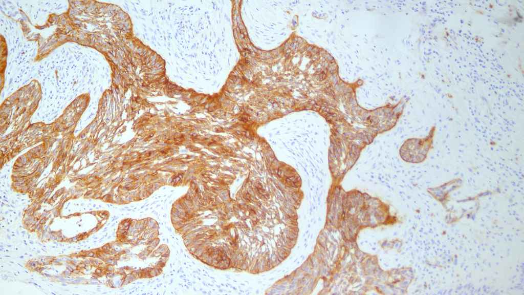
If you think of modern oncology as Sherlock Holmes, then consider IHC its trusty magnifying glass—vital for cracking complex cases of cancer diagnosis and treatment plans. Here’s why: thanks to special stains used during IHC detection systems such as DAB substrate kits or chromogenic detection involving alkaline phosphatase, we can now color-code cells suspected of foul play amid healthy neighbors.
This color-coding party gets even more exciting when multiple epitopes are involved because now there are different shades highlighting multiple antigens simultaneously—and who said science wasn’t vibrant? What makes all this possible is selecting primo primary antibodies which latch onto target antigen areas while secondary antibodies work double-time amplifying signals so not even the sneakiest protein goes undetected.
Did you know? The accuracy range for diagnosing cancers using successful IHC runs between 70% to 90%. That means when tissues get tagged with an antibody detective squad searching high-and-low under microscopes—we’re winning most battles against misdiagnoses.
Principles and Methodologies of Immunohistochemistry Staining
Dive deep into what makes your body tick—or rather stick—in terms of cell chatter through immunostaining methods like antibody binding games happening right beneath our noses…or skin layers.
Principles and Methodologies of Immunohistochemistry Staining
Peering into the heart of tissues to see where specific proteins hang out is no small feat. That’s where immunohistochemistry (IHC) staining comes in, not just as a fancy technique but as an essential partner for scientists wanting to spot that elusive target antigen. Think of IHC like a high-stakes game of hide and seek, with antibodies being your seasoned seekers.
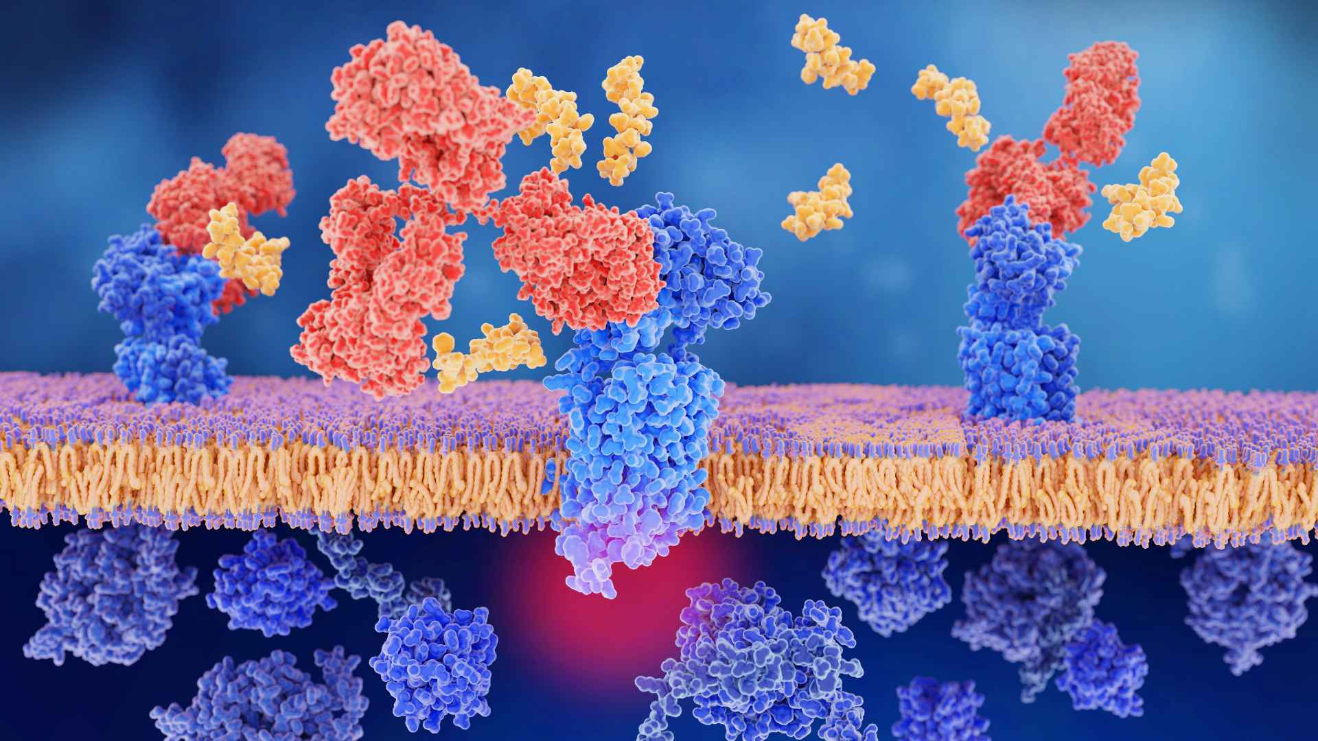
Antibody-Antigen Specificity
The power couple at the core of this technique are antibodies and antigens – it’s all about compatibility here. The primary antibody has one job: find and bind its matching antigen within the tissue sample like they’re long-lost soulmates. This isn’t some random match-making; it’s precision down to the molecular level because specificity matters when you’re looking for that needle in a biological haystack.
Pick your player wisely though—primary antibodies come from various host species, each bringing their own strengths to the table. You might get lucky with polyclonal antibodies snagging multiple epitopes on your target or play it cool with monoclonal ones that know how to zero in on just one special stain.
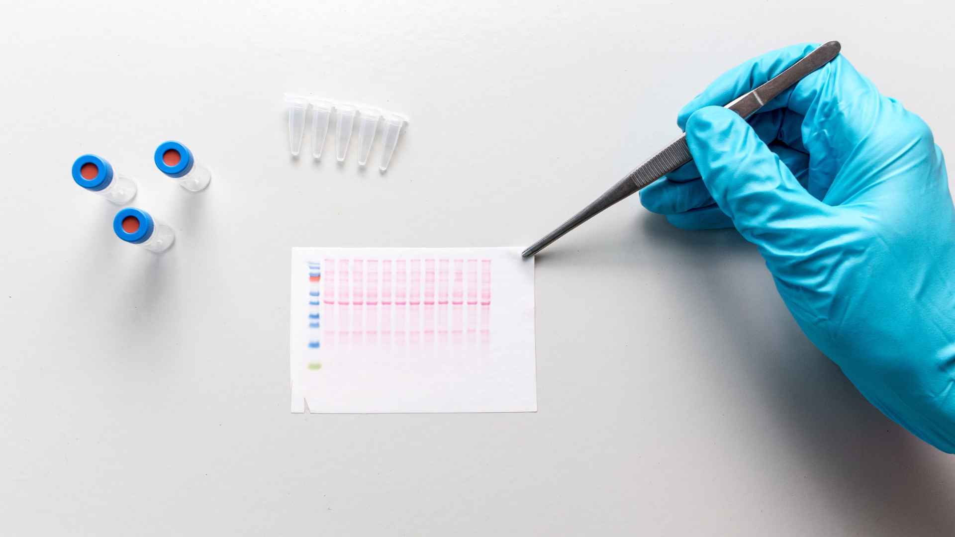
Visualization Techniques
We’ve tagged our targets; now let’s light them up. Enter stage left: secondary antibodies carrying flashy labels like horseradish peroxidase or alkaline phosphatase – enzymes known more for their visual flair than any culinary prowess. These aren’t merely accessories but critical pieces for signal amplification so you can actually see what’s going down under the microscope.
If we want even more drama, chromogenic detection methods bring color into play using substrates such as DAB substrate kits which turn brown when they react — because who doesn’t love a bit of color-coded clarity? But sometimes we crave subtlety; then fluorescent labeling takes over making everything glow if UV lights hit just right – think neon signs pointing straight at our protein pals.
The Evolution and Impact of Immunohistochemistry Staining
Casting our gaze back through time reveals IHC wasn’t always this sophisticated darling tool—it was born out of necessity during battles against diseases unknown back then . Who knew pneumonia had bacterial culprits until someone thought to dab those samples with dyes?
Pioneering Moments in IHC
The year 1941 marked history books—not only were conflicts brewing worldwide but also within labs leading us towards scientific breakthroughs. The first successful IHC stain set off fireworks, signaling hope amidst darker times by revealing details never seen before.
Antigen Retrieval Techniques in Immunohistochemistry Staining
Tissue samples are like time capsules, preserving the secrets of cellular interactions. But sometimes, during tissue fixation, these biological narratives get a little scrambled—epitopes hide away as if playing an intense game of hide-and-seek with researchers. Antigen retrieval techniques in immunohistochemistry (IHC) staining are like the skilled seekers who coax them out into the open.
Heat-Induced Epitope Retrieval: The Spa Treatment for Tissues
The first method we’ll explore is heat-induced epitope retrieval (HIER). Imagine giving your tissues a spa day where steam baths help relax and reveal those elusive antigens that got cozy under cross-linking proteins during formalin fixation. It’s not just any hot spring dip though; it’s about precision heating usually involving citrate or EDTA buffer solutions to break those protein bonds without damaging tissue architecture.
This technique has proven so effective that it’s become one of the two main methods used by pathologists and scientists alike when they want to ensure their IHC data shines bright with clarity.
Enzymatic Retrieval: When You Need Just a Pinch More Precision
Sometimes what you need isn’t more heat but rather a targeted approach—that’s where enzymatic retrieval steps onto the scene. Picture tiny molecular scissors snipping at proteins selectively, unveiling antigenic sites with surgical precision while keeping delicate tissue morphology intact. Enzymes such as trypsin or pepsin gently dissect intermolecular forces ensuring antibody binds specifically and firmly to its target antigen.
If HIER is akin to taking down walls to find something lost behind them, then think of enzymatic retrieval as picking locks for a less destructive revelation—the choice between them often depends on which key fits best for unlocking your particular ihc study mystery.
Da-Ta Biotech L.T.D.’s focus areas, dive deep into optimizing these methodologies because let’s face it; nothing beats experience when you’re trying to improve IHC staining quality.
Finding Your Method: Matchmaking Antigens With Their Ideal Technique
You wouldn’t use dynamite fishing if you’re looking after tropical fish in an aquarium—and similarly, selecting between HIER or enzymatic methods should be done considering factors like antigen sensitivity and required detection thresholds. For instance, some might say applying too much heat can turn our specimen ‘soup’—but knowing exactly how much temperature infusion works best comes from practical know-how gained through countless trials across multiple antigens and sample preparations within Da-Ta Biotech’s labs.
- To assess if you meet the criteria for our program, we will carefully examine your application.
Selecting Antibodies for Optimal Immunostaining Results
Imagine you’re a detective on the trail of cellular culprits, and your leads are as tangled as headphone wires in your pocket. Selecting the right antibodies for immunohistochemistry (IHC) staining is akin to choosing the perfect magnifying glass—it can make or break your case. Let’s crack this wide open.
The Quest for Specificity: Primary Antibody Selection
When it comes to primary antibodies, specificity is king. Think of these like heat-seeking missiles locked onto their target antigen with laser focus. They need to bind precisely without latching onto innocent bystanders—aka non-target antigens—to avoid misleading background noise that muddles our IHC data.
To achieve this Sherlock-Holmes level of deduction, consider factors such as whether monoclonal or polyclonal antibodies fit the bill. Monoclonals are steadfast; they recognize a single epitope—a specific part of an antigen—with unwavering loyalty but may miss out if that very epitope is obscured post-tissue fixation. Polyclonals play wider fields and bind multiple epitopes, raising detection odds even when some targets are masked by formalin’s tight grip during tissue preparation.
You’ll also want to ensure compatibility between antibody species and host species—after all, cross-reactivity could lead us astray faster than false rumors in high school hallways. So take time selecting wisely from databases listing well-characterized options.
The Supporting Act: Secondary Antibodies Amplify Signals
Your secondary antibody should be loyal—not just any lab partner will do here. This tag-team player binds directly to primaries while carrying markers like horseradish peroxidase or alkaline phosphatase which act like amplifiers at a rock concert—turning up volume so we can hear every note clearly through chromogenic detection using substrates such as DAB substrate kits.
Bear in mind though; too much signal amplification via methods like ABC method can backfire worse than mic feedback—that pesky background staining dab won’t go unnoticed. Thus achieving balance between visibility and accuracy demands finesse—the kind required when seasoning food just right.
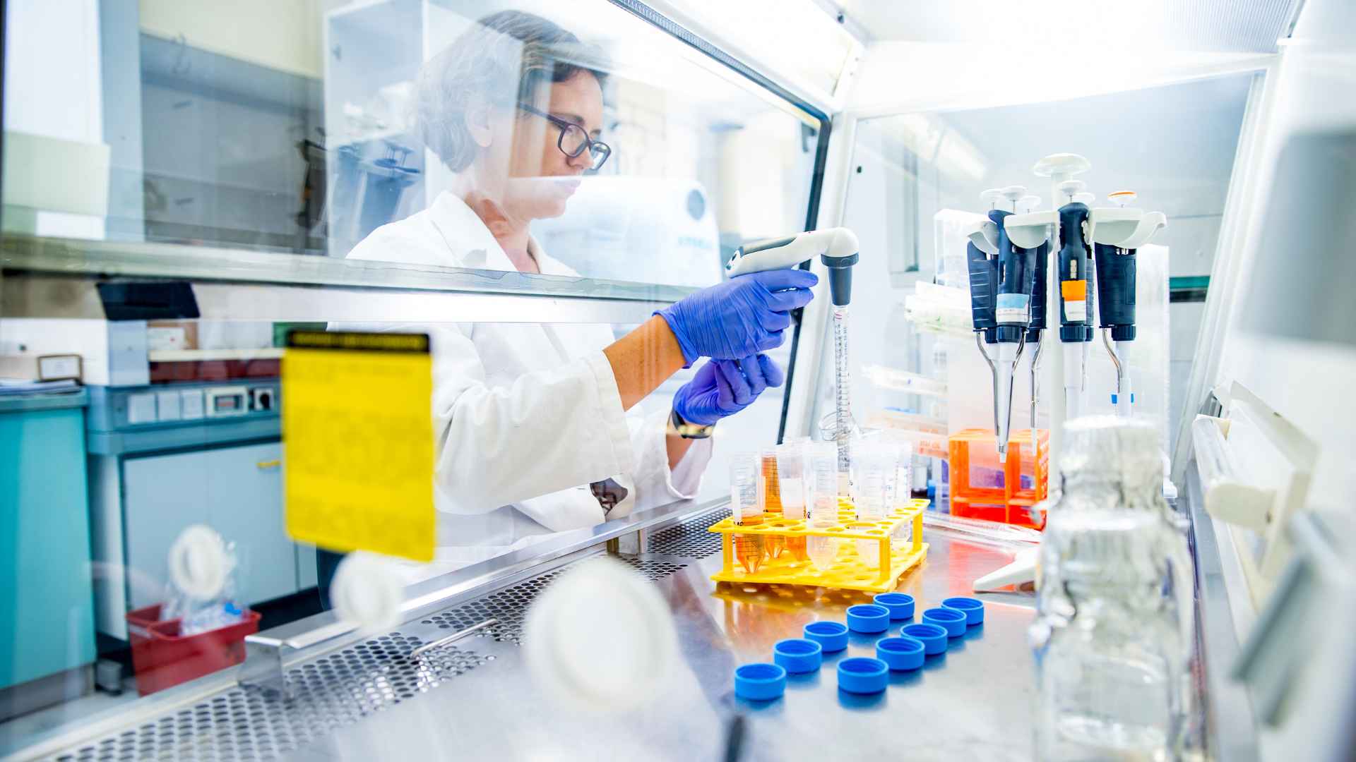
Tailoring Tissue Preparation Techniques For Success
To ensure a successful IHC study, meticulous attention to sample preparation is critical. This means careful tissue collection, proper embedding techniques, and sectioning that prevents ice crystal formation which can damage the intricate structures within frozen samples. It’s also vital to avoid common staining errors like those caused by Hoechst dye misapplication that can lead to misleading nuclear blue artifacts.
Conclusion
Start by painting with precision. Immunohistochemistry staining lets you trace diseases, one cell at a time. Remember the pioneers who brought us here and how today’s cancer diagnosis hinges on this craft.
Think specificity; your antibodies are your scouts, seeking out target antigens like detectives on a mission. Know that each antigen retrieval method unveils hidden truths in tissue architecture.
Pick wisely; selecting primary and secondary antibodies shapes the clarity of your data generated. Each choice amplifies signals, drowning out noise to detect expressed antigens with finesse.
So stand firm in knowledge—knowing every stain tells its own vital story—and let immunohistochemistry guide you to new discoveries beneath the microscope’s gaze.
Finde out more about our research services by clicing our home page.